Retinal Detachment Surgery
Retinal detachment is a sight-threatening emergency requiring urgent surgery from a specialist vitreoretinal surgeon to prevent permanent vision loss


Excellent
4.96 out of 5
Overview
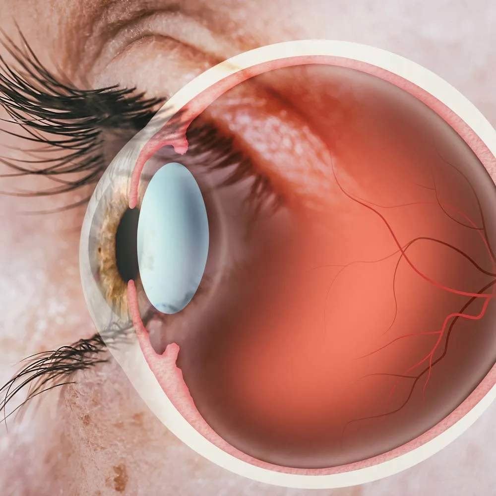
A retinal detachment is when the retina comes away from the wall of the eye causing loss of sight. The retina is like the film of a camera, and the longer it remains detached, the more damage that occurs. For this reason, a retinal detachment is an emergency scenario which in the vast majority of cases requires urgent retinal detachment surgery. The sooner the retina is reattached, the better the long term vision. In most cases, the operation of choice is called a vitrectomy.
In the majority of cases, patients will notice symptoms such as new floaters, flashing lights and a black curtain coming across their vision. However, not all patients have all symptoms, and occasionally there might be very minimal symptoms occuring. Some patients describe the flashes as being like an arc of light in the side of their vision, but again, this can vary on a case-by-case basis. Whenever there are symptoms such as flashes, floaters or a shadow/curtain in the vision, it is important to be assessed urgently by a retinal specialist such as myself. In many cases, there may not be a suspicious cause. However, early diagnosis of a retinal detachment allows for urgent treatment and ultimately better long-term vision.
The urgency of surgery depends on the type and severity of the retinal detachment. Terms such as ‘macula-on’ or ‘macula-off’ are often mentioned. The macula is the part of the retina which is responsible for central vision. A ‘macula-on’ retinal detachment is a term used when the detachment is only affecting the peripheral retina and therefore the central vision can often be relatively preserved. In these emergency cases, it is important to reattach the retina as soon as possible and we usually aim for this to be done within 24 hours of diagnosis, depending on availability of surgical theatres. Macula-off retinal detachments also need to be repaired urgently. In these cases, fluid had already collected under the macula and this means damage is occurring to the photoreceptors (light sensing cells). The longer it takes to reattach the retina, the more the damage that occurs and this means less recovery of vision after surgery. In practical terms, this can potentially be the difference between being able to retain a driving license, or losing it.
Causes & Risk Factors
A retinal detachment can happen to anyone at any age. However, there are certain people who are more at risk.
The most common age group for a retinal detachment is 50-70. This is because it is the period when the vitreous gel (jelly) inside the eyeball undergoes changes which result in it shrinking away from the back of the eye. This is known as a posterior vitreous detachment and I have written a previous blog about this. During a posterior vitreous detachment, a retinal tear can occur. A retinal tear is a major risk factor for developing a retinal detachment and this is why anyone with symptoms of a posterior vitreous detachment should have their eyes examined urgently by an expert ophthalmologist.
A major risk factor for developing a retinal detachment is myopia (being short sighted). This is because the eyeball is usually bigger in myopic patients, and therefore the retina is thinner since it is stretched over a larger surface. In addition, myopic patients undergo a posterior vitreous detachment (often the event that precedes a retinal detachment) earlier than normal sighted or long-sighted patients. It is also very important to be aware that patients who have had laser eye surgery to correct short sightedness still have this increased risk of developing a retinal detachment.
Another risk factor to be aware of is recent eye surgery, for example cataract surgery. We also know patients who suffer a retinal detachment in one eye are at a significantly increased risk of developing a retinal detachment in their other eye. Other risk factors include trauma to the eye, and in rare cases eye tumours.
Diagnosis
A retinal detachment is a clinical diagnosis. This means it is diagnosed by examining the eye at a specialist microscope and directly looking at the retina through the pupil. Best practice is to use dilating eye drops to make the pupil larger and ensure a careful and accurate assessment of the retina is possible. Seeing a retinal surgeon like myself is a good idea if you are concerned about a retinal detachment. This is because we will usually perform other specialist examination techniques, such as indenting the edge of the eye, in order to fully visualise the entire retina. Furthermore, seeing a retinal surgeon saves time because if a retinal detachment is diagnosed, we are in a position to rapidly arrange a surgical procedure to repair it.
During your examination, you may also be recommended to have an OCT scan of your retina. This is a painless and quick scan (less than 30 seconds) which provides helpful information about the severity and type of retinal detachment.
Examination of the eye is a painless procedure. It is important to be aware that you cannot drive for 4-6 hours after dilating drops, however. For these reasons, thinking about travel arrangements is a sensible idea.
Operations & Procedures
There are two main surgical procedures for retinal detachment.
Firstly, a vitrectomy is a microsurgical minimally invasive keyhole surgical procedure that is appropriate for the vast majority of retinal detachment surgery. This operation can be performed under local anaesthetic, sedation or general anaesthetic and takes approximately 60 minutes. This operation involves using a high speed cutter (10-30,000 cuts per minute) to remove the vitreous gel (jelly) from inside the eyeball. The retinal detachment is then repaired by draining the fluid from underneath the retina and using a laser or cryo (freeze) probe to repair any retinal tears which have caused the retinal detachment. A gas bubble is then usually inserted into the eye to help with recovery and improve the likelihood of surgical. Less commonly, an oil bubble can be needed.
Another option for retinal detachment repair is by using a scleral buckle. This is where a small piece of plastic is sutured to and hidden within the wall of the eye. It is an older technique, but remains the operation of choice for a small subset of retinal detachment types. This operation usually requires a general anaesthetic.
When I examine your eye and diagnose a retinal detachment, I will explain to you which type of surgery I recommend and the risks and benefits.
What to Expect During Your Surgery and Why Choose Mr Neffendorf
Retinal detachment surgery usually needs to be scheduled urgently, often within 24 hours. My team will ensure this process is as stress-free as possible because we understand how stressful this scenario is for patients. We will ensure you are well informed about your procedure and make sure you know where and when to report.
On arrival to the hospital, you will be greeted by the nursing staff and taken to a room where general checks can take place, for example measuring your blood pressure. I will then come to see you to talk about the operation and answer any remaining questions you may have. If you have chosen to have sedation or a general anaesthetic, this will also be a time when you can speak to the anaesthetist.
The surgery itself takes approximately 1 hour. There should not be any significant pain during the surgery. If you have chosen a local anaesthetic, you may see some lights from the operating microscope during the surgery or some patterns of coloured lights. However, you will not see the actual operation through the eye that is being operated on, nor from your other eye which will be lightly covered by a drape.
I will talk to you during your operation to keep you relaxed and you can let me know at any time if you feel pain. This will allow me to top-up your anaesthetic and ensure you are comfortable.
Recovery
Once you have recovered after the surgery and are feeling comfortable, you will be able to leave hospital. Depending on your anaesthetic type, you may feel a little tired, and therefore it is important to have a relative or friend with you to assist with your journey home. There will be a pad and shield over the eye. I will explain when these are to be removed.
You will be provided with eye drops to take and these are started on the day after surgery. These contain antibiotic and steroid and are to reduce the chance of developing an infection as well as help with the healing process. There may be some mild ache in the eye which can be managed with simple painkillers such as paracetamol or ibuprofen. Significant pain is uncommon.
I will have provided you with my contact details and information about what to do if you have any concerns. In general, the signs to look out for in the early post-operative period are significant or worsening eye pain, nausea and vomiting, a painful red eye or substantial loss of vision. Most patients will have a gas bubble in the eye after surgery and this means the vision will be very blurred when the pad is removed. Often patients describe it as feeling as though they are underwater.
Time off work varies from patient to patient and is dependent on the nature of the surgery as well as the type of work you do. In general, I advise 2 weeks off work. It is also not advisable to drive for a few weeks following surgery, but I will have discussed this with you both before and immediately after surgery.
In order to improve the vision after surgery, the best thing is to follow the post-operative instructions. This will maximise the chances of a successful retinal reattachment, and best vision.
The ‘Gas Bubble’ & Posturing
Patients often come to an appointment with questions about the gas bubble as well as the need for posturing. There are different types of gases, and they take varying amounts of time to be absorbed by the body.
In most retinal detachment operations, one of the final steps is to insert a gas bubble into the eye. This is to maintain a pressure and help increase the chances of successful retinal reattachment. The gas bubble pushes against the retina to help it stick back in place. Depending on the type and concentration of gas used, it takes anywhere from 4 weeks to 3 months to fully absorb. Whilst gas is inside the eye, you cannot fly in an airplane or be at altitude due to the pressure changes that occur.
The rationale for posturing is to move the bubble into a position where it will be most effective in helping the retina to stay attached. Posturing varies for different patients, but in the case of retinal detachment surgery, face down is not usually used. More commonly, we might ask a patient to lie on one side as much as they can for a week, or to stay upright (and sleep propped up with a few pillows). Most patients find this acceptable and not too difficult to do. Posturing can be challenging, but we only ever ask for a maximum of 7 days.
Success Rates and Long-Term Outcomes
For most retinal detachments, the rate of successful retinal reattachment is 80-90% with one operation. If surgery is unsuccesful and the retina doesn’t reattach, we can perform more surgery which carries a good chance of repairing the detachment. There are certain scenarios where the success rate is lower. Often this is when a detachment is diagnosed late in a more advanced stage, emphasising the importance of early recognition and assessment by a retinal specialist in the event of retinal detachment symptoms (see above).
If you suffer a retinal detachment in one eye, you are statistically more likely to develop a retinal detachment in your other eye compared to someone who has never had a retinal detachment. In some cases, there may be preventative measures that can be done to reduce this risk, and I will discuss whether these apply to you.
Symptoms
In the majority of cases, patients will notice symptoms such as new floaters, flashing lights and a black curtain coming across their vision. However, not all patients have all symptoms, and occasionally there might be very minimal symptoms occuring. Some patients describe the flashes as being like an arc of light in the side of their vision, but again, this can vary on a case-by-case basis. Whenever there are symptoms such as flashes, floaters or a shadow/curtain in the vision, it is important to be assessed urgently by a retinal specialist such as myself.
Surgery
Surgical treatment for retinal detachment
Vitrectomy

This operation involves using a high speed cutter (10-30,000 cuts per minute) to remove the vitreous gel (jelly) from inside the eyeball. The retinal detachment is then repaired by draining the fluid from underneath the retina and using a laser or cryo (freeze) probe to repair any retinal tears which have caused the retinal detachment. A gas bubble is then usually inserted into the eye to help with recovery and improve the likelihood of surgical. Less commonly, an oil bubble can be needed.